Did you know that one in eight women will develop an invasive breast cancer in her lifetime? Your risk is not reduced just because breast cancer does not run in your family. Knowing this, the industry has evaluated using breast thermography vs mammogram, and you should be aware of the differences.
Something has to change
In order to decrease the high incidence of breast cancer, we must re-evaluate our current screening procedures. Breast thermography is a safe and highly sensitive screening tool for early detection of breast cancer. 1
How breast thermography works
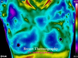
Thermography uses infrared technology to detect minute changes in the breast tissue by analyzing vascularity (blood flow) in the breast. Healthy breasts should appear symmetrical, and vascular patterns and temperature should be relatively the same from one breast to the other.
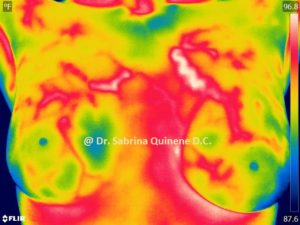
 Asymmetrical vascularity, or differences in heat from one breast to another, can indicate a potential risk factor for developing breast cancer. The photo on the left is an example of an individual with increased risk. Both breasts show hypervascularity which is more pronounced in the left breast.
Think of breast thermography like a thermometer. When a child first develops a fever, a mother checks her temperature with a thermometer to detect for illness. It may not be clear what illness is present when there is a fever—it could be anything from the common cold to something as severe as meningitis. But the fever is a warning sign that the body is fighting a disease.
When a “fever” or temperature difference in the breast is detected, the cause may still be unknown, but it is an early indication that the body is combating something foreign.
Thermography detects earlier than mammograms
To understand the main benefit of thermography—early detection—we must first understand how cancer works. Every time a cell in the body divides there is an opportunity for an abnormal mutation. Substances like toxins in the foods we eat and the air we breathe, or free radicals we are exposed to, can be dangerous to our DNA replication process.
A cell doubles every 90 days. When the cell is malignant, the following numbers show the rate at which a tumor develops.
- day 1 = 1 malignant cell
- 90 days = 2 malignant cells
- 1 year = 16 malignant cells
- 3 years = 4,096 malignant cells
- 4 years = 65,536 malignant cells
- 5 years = 1,048,576 malignant cells (Even at 5 years, breast cancer is usually still undetectable)
- 6 years = 16,777,216 malignant cells (It is not until years 6 through 8 that cancer will become apparent)
Waiting until year six when a mammogram can detect cancer creates a significant time lapse between onset, diagnosis, and treatment. During this time, the tumor can be transforming from benign to malignant and the prognosis might suffer as a result.
Depending on the size and density of the breast, a mammogram might not be able to detect a tumor until it is the size of a pea (after six to eight years of developing). A thermogram can detect an abnormality during the first few years, while it might not necessarily be cancer, and therefore provide the best chances for treatment.
Advantages of thermography
Unlike mammograms, thermograms do not involve radiation. It’s quite counterintuitive to radiate the breast when radiation can be a contributing factor to the development of cancer.

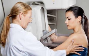
Mammograms compress your breast tissues and can be very painful
Unlike mammograms, where the machine can add an equivalent of 50 pounds of pressure to the breast tissue, thermography involves no contact. Therefore, thermograms are great for those with implants, expectant moms, and women with tender breasts.
Mammograms are known to produce many false positives, meaning there is a high percentage of diagnosed cancer that might not necessarily be cancerous. This leads to unnecessary psychological stressors, an economic burden, and further procedures such as biopsies and mastectomies.
According to the British Medical Journal, annual mammography performed on women ages 40 to 59 does not reduce mortality from breast cancer beyond that of physical examination. Therefore, mammograms have not been instrumental in decreasing the mortality rate. 2
Unlike mammography, breast thermography images the entire chest including the axilla (underarm). This is important because lymph drains from your armpit into the breast tissue. Many cancers are found in the upper outer quadrant where the lymph drains from, so it is very important to include this area in the thermogram. A mammogram only takes a picture of the breast tissue that can fit in the machine and be compressed.
Thermography vs mammogram which is better?
The best data in terms of saving lives comes when multiple imaging modalities are performed, providing a better, more complete picture of the breast. For this reason, thermography is not a replacement for a mammogram but is used as an adjunct screening procedure for the early detection of cancer.
Thermograms should be used beginning at the age of 25 when the breasts are fully developed, to provide a good baseline to contrast against any future abnormalities.
 Many physicians perform breast thermography, but unfortunately, some are performing thermography inadequately. Make sure that your physician is taking not only color images, but black hot images to show the defined vascularity. It is also important that your doctor has a defined grading scale for different levels of vascularity. The breast thermogram in the photo to the left is an example of hot images showing hypervascularity.
To find a physician in your area that performs thermography, visit the Women’s Academy of Breast Thermography. You can gain peace of mind through safe and early breast thermography detection for you and each of your loved ones.
__________________________________________________
1 Efficacy of Computerized Infrared Imaging Analysis to Evaluate Mammographyically Suspicious Lesions. American Journal of Radiology, January 2003.
2 Twenty-Five Year Follow-up for Breast Cancer Incidence and Mortality of the Canadian National Breast Screening Study: Randomised Screening Trial. Hwadmin. British Medical Journal, 11 Feb. 2014. Web. 24 Feb. 2015.


